Radiology
Section outline
-
Welcome to Children’s National Hospital (CNH) and the Department of Radiology!
CNH was ranked No. 5 nationally in the U.S. News & World Report 2022-23 Best Children’s Hospitals annual rankings. This marks the sixth straight year Children’s National has made the Honor Roll list, which ranks the top 10 children’s hospitals nationwide. In addition, its neonatology program, which provides newborn intensive care, ranked No.1 among all children’s hospitals for the sixth year in a row.
Our pediatric radiology residency training program provides a plethora of clinical experience in all areas of pediatric radiology, including teaching, research, administrative activities in the areas of quality assurance, organization, and management.
Our 21 board certified Radiologist work very closely with residents and fellows in our state of the art facility. We hope you have an enjoyable learning experience at Children's National!
Our imaging and radiology services include:
· CT (computerized tomography)
· Neuroimaging
· Cardiac imaging
· Nuclear medicine
· DEXA-scan (bone scan)
· Orthopaedic and Rheumatology imaging (MSK)
· Diagnostic X-rays
· PET scan (positron emission tomography)
· Fetal MRI and US
· Ultrasound (US)
· Fluoroscopy
· Interventional Radiology (IR)
· MRI (Magnetic Resonance)
· Neuro-Interventional Radiology (NIR)
-
On behalf of the faculty of Children's National Hospital, we would like to welcome you to the training program in pediatric radiology. Our training program provides a wide variety of experience and teaching in all of the areas of pediatric radiology.
MISSION: Our mission is to be the preeminent provider of quality health care services to infants, children and youth in our region. Children's National will strive to enhance the health and well-being of all children through responsible programs of excellence in research, education and advocacy.
Our pediatric training mission is to train individuals who will provide high quality imaging care of children and who will serve as leaders and mentor in responsible practice, in education, in research and in advocacy.
VALUES: Each trainee in pediatric radiology is encouraged to adopt the values of Children's National. These include provision of excellence in clinical service, patient care, research, advocacy and education. Compassionate treatment of children and their families is paramount. Development of outstanding relationships with colleagues, co-workers and referring physicians is essential.
Before you can begin your rotation, all paperwork must be completed in MedHub. As a reminder, paperwork is renewed every academic year.
-
-
-
-
-
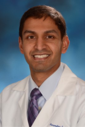 Dr. Narendra Shet
Welcome to the exciting world of Body MR! This rapidly evolving field can seem daunting at times but has a plethora of applications in kids and is particularly appealing since MR is radiation-free. For example, we can use dynamic MR to perform multiphase evaluation of a lesion, to know whether it enhances in the arterial, portal venous, and/or late phases; multiphase CT, while sometimes used in the adult population, carries a lot of potential risks to the developing pediatric body. We can use MR to evaluate for a wide variety of structures and organ systems; for example, we can use MR to evaluate pediatric abdominal masses, but we can also use MR in the acute setting to assess for appendicitis or osteomyelitis. We can optimize MR to evaluate structures that might have poor intrinsic signal on MR, such as by filling the bowel with fluid during MR enterography in order to get rid of air signal and better visualize mural architecture and enhancement.
While there are quite a few things to be learned on the MR rotation, the overarching goals are to:
- Be a consultant for referring clinicians and direct them toward the right test (and right modality – sometimes MR is not the best choice!)
- Learn the basics of MR physics and sequences, and choose the right protocol for an exam
- Understand normal and abnormal appearances of structures on MR, depending on the sequence
- Create accurate and succinct reports for clinicians, using Powerscribe templates as a basic framework
It may be difficult to achieve goals #1-3 for rotating residents due to limited time on the MR service, but we will do our best to introduce you to pediatric body MR and spark your interest in spending more time with us! Since fellows have a more longitudinal experience on the service, the following goals/objectives are expected to be achieved at various timepoints.
Quarterly Goals/Objectives:
July-September:
- Develop understanding of workflow on CT/MR rotation – what studies are to be read
- Learn how to protocol in Radnet with supervision, including how to order contrast
- Learn basic protocol options for commonly encountered body MR studies (e.g., MR enterography)
- Learn how to open MR study in PACS, including various series
- Contact IT if you would like assistance in adjusting your hanging protocols
- Go over what to look for when doing QA for a study
- Know the MR appearance of various normal organs on common pulse sequences such as T2 and know the classic appearance of acute abnormalities such as appendicitis
- Draft reports for MR studies using Powerscribe templates
October-December:
- Protocol studies in Radnet, at the start of the workday and when contacted by MR technologists
- With guidance from your attending, know when to select gadolinium-based contrast agents other than Dotarem
- Select protocols for commonly encountered body MR studies (e.g., MR enterography) in Radnet
- Perform study QA for common body MR studies when contacted by MR technologists, with attending guidance
- Know the MR appearance of various normal organs on common pulse sequences such as T2 and characterize acute and non-acute abnormalities
- Draft reports for MR studies using Powerscribe templates; be sure to include correct sequences used (these may differ from template)
January-March:
- Protocol studies in Radnet, at the start of the workday and when contacted by MR technologists
- Select protocols in Radnet for commonly and uncommonly encountered body MR studies
- Perform study QA for common and uncommon body MR studies when contacted by MR technologists, with decreasing attending guidance (if OK’d with your attending, can perform QA independently, but attending should notify technologist that fellow QA is acceptable, to avoid confusion)
- Know the MR appearance of various normal organs on common pulse sequences such as T2 and characterize acute and non-acute abnormalities
- Review uncommon body MR studies such as whole-body MRI
- Draft reports for MR studies using Powerscribe templates; be sure to include correct sequences used (these may differ from template)
April-June:
- Protocol studies in Radnet, at the start of the workday and when contacted by MR technologists
- Select protocols in Radnet for all body MR studies
- Perform study QA for common and uncommon body MR studies when contacted by MR technologists, with little attending guidance
- Know the MR appearance of various normal organs on common pulse sequences such as T2 and characterize acute and non-acute abnormalities
- Review any body MR studies encountered while on service
- Draft reports for MR studies using Powerscribe templates; be sure to include correct sequences used (these may differ from template)
Resources:
Cases:
a. In PACS, under username nshet, cases under Teaching Cases/Body MR
Textbooks:
a. The Physics of Clinical MR Taught Through Images – Runge, Nitz, Schmeets (2005)
b. Pediatric Body MRI: A Comprehensive, Multidisciplinary Guide – Lee, Liszewski, Gee, Daltro, Restrepo (2020)
· If you have access, use this resource; however, the field is so quickly developing that articles are often the most up to date and accurate for clinical practice.
a. MR Physics, Sequences, and Techniques: ·
Moore MM, Chung T. Review of key concepts in magnetic resonance physics (2017)
Jo S et al. Musculoskeletal MRI Pulse Sequences: A Review for Residents and Fellows (2019)
Jaimes C et al. Fast, free-breathing and motion-minimized techniques for pediatric body magnetic resonance imaging (2018)
Kozak BM et al. MRI Techniques to Decrease Imaging Times in Children (2020)
b. MR Contrast: ·
Ayyala RS et al. Intravenous gadolinium-based hepatocyte-specific contrast agents (HSCAs) for contrast-enhanced liver magnetic resonance imaging in pediatric patients: what the radiologist should know (2019) ·
Blumfeld et al. Gadolinium-based contrast agents — review of recent literature on magnetic resonance imaging signal intensity changes and tissue deposits, with emphasis on pediatric patients (2019) ·
Farmakis SG et al. Safety of gadoterate meglumine in children younger than 2 years of age · Ponratana S et al. Safety issues related to intravenous contrast agent use in magnetic resonance imaging (2021)
c. MR Findings/Interpretation: ·
Moore MM et al. MRI for Clinically Suspected Pediatric Appendicitis: Case Interpretation (2014) ·
Mollard BJ et al. Pediatric MR Enterography: Technique and Approach to Interpretation--How We Do It (2015)
Chan, BY et al. MR Imaging of Pediatric Bone Marrow (2016) ·
Navarro OM et al. Pediatric Soft Tissue Tumors and Pseudotumors: MR Imaging Features with Pathologic Correlation. Part 1. Imaging Approach, Pseudotumors, Vascular Lesions, and Adipocytic Tumors (2009) ·
Pediatric Soft Tissue Tumors and Pseudotumors: MR Imaging Features with Pathologic Correlation. Part 2. Tumors of Fibroblastic/Myofibroblastic, So-called Fibrohistiocytic, Muscular, Lymphomatous, Neurogenic, Hair Matrix, and Uncertain Origin (2009) ·
Shet NS et al. Use of Eovist in Pediatric Patients: Pearls and Pitfalls (2020) ·
Dickerson EC et al. Pediatric MR Urography: Indications, Techniques, and Approach to Review (2015) ·
Gottumukkala RV et al. Current and Emerging Roles of Whole-Body MRI in Evaluation of Pediatric Cancer Patients (2019) ·
Acharya PT et al. Pediatric Mediastinal Masses: Role of MR Imaging as a Problem-Solving Tool (2019)
d. MR Safety/Quality · Artunduaga M et al. Safety challenges related to the use of sedation and general anesthesia in pediatric patients undergoing magnetic resonance imaging examinations (2021)
-
-
-
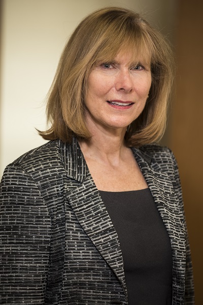 Dr. Dorothy Bulas
Welcome to Fetal Imaging.
Pediatric Radiology includes the exciting field of “Fetology”. Imaging of the fetus is fascinating in many ways, helping us understand the pathophysiology of numerous abnormalities, as well as the normal growth and development of the fetus. As a fetal imager, one works with numerous subspecialties to help determine appropriate perinatal care, and postnatal management. Counseling of families is a moving experience, with multiple medical and ethical issues to consider. Much research still needs to be done with advances in fetal imaging – Doppler, 3D/4D, MR spectroscopy, DWI/EPI helping advance fetal intervention.
Ultrasound is the modality of choice when imaging the pregnancy and fetus. It is noninvasive, safe due to absence of radiation, low in cost and has widespread availability. The technique has high accuracy and superior spatial resolution, allowing real time, color Doppler, multiplanar and 3-4 dimensional capabilities.
When an abnormality on US is not clearly defined and more information is sought in order to decide about therapy, delivery, or to advise a family about prognosis MR can be performed. MR can improve the assessment of a potential anomaly particularly in the setting of maternal obesity or oligohydramnios. Fetal MR is particularly powerful in the evaluation of the fetal brain and can help asses lung volumes in cases of congenital diaphragmatic hernia or lung masses.
Fetal Imaging Rotation Tips:
Fetal cases are scheduled Tuesday through Friday.
MR cases start at 7 AM with US to follow through the early afternoon.
Have the fetal center send you the case list the day prior to review the histories and read up on the diagnosis.
Notify the fetal attending each day you are on fetal so they expect you, know where to locate you and can start reviewing cases with you in real time if appropriate.
Atleast a few times during your rotation, skip a 7 30 didactic conference and go sit with the MR techs and watch fetal MR scans being performed.
Atleast a few times during your rotation, sit with the sonographer and watch an OB US completed – learn how to accurately measure HC/BPD/FL/AC.
Discuss with the attending which fetal MR and OB US cases to dictate – templates available
Atleast a few times, arrange with the fetal neuroradiologist to review fetal MR neuro cases together. Ask if you can dictate a neuro case.
Participate in the daily 11 AM case review
Take advantage of the Prenatal Pediatric Institute which is a consortium of subspecialists throughout the hospital with a particular interest in fetal care. Weekly meetings and numerous CME lectures are offered throughout the month.
Attend Weekly Fetal Meeting - Tuesday from 7 – 8 AM
Attend CME lectures – usually from 12-1 Mondays and Tuesdays monthly


PPI Ethics Rounds
Hundreds of fetal cases can be reviewed under Synapse All users/bulas,dorothy/fetal…..
Fetal competencies
Participate fully on service
Watch Fetal MR techs scan
Watch OB sonographers scan
Review fetal case histories the night before
Read about the diagnosis to be imaged the night before – pull articles or read the appropriate chapter in Fundamental and Advanced Fetal Imaging
Review cases with the fetal body and fetal neuro attendings each day.
Learn normal fetal brain anatomy for each GA via the atlas in Fundamental and Advanced Fetal Imaging
Dictate select fetal body and neuro cases
Present a case each week in interesting case conference
Consider working on a poster or abstract on fetal imaging
Recommended Reading:
Fundamental and Advanced Fetal Imaging: US and MRI Kline-Fath, Bulas, Lee (2021) Wolters Kluwer
MRI of the Fetal Brain: Normal Development and Cerebral Pathologies. C Garel et al; Springer (2004).
Fetal MRI. D Prayer (ed.), Springer (2011)
Mini-symposium on Fetal MRI. Pediatric Radiology, volume 34(9): 681-719. (2004) http://www.springerlink.com/content/0301-0449/34/9/
Fetal magnetic resonance imaging: jumping from 1.5 to 3 tesla T Victoria et al Pediatr Radiol (2014) 44:376–386 DOI 10.1007/s00247-013-2857-0
Web sites:
SPR Fetal MRI web site – filled with numerous educational posters, protocols, templates: https://www.pedrad.org/Specialties/Fetal-Imaging/Fetal-MRI-General-Information
Twitter: @fetal_imaging
Instagram username – fetal_imaging
Facebook - https://www.facebook.com/fetal.imaging
http://www.fetalmedicine.com - US education, including videos of diagnoses
http://www.thefetus.net - educational material
http://www.ob-ultrasound.net/ - multiple links to OB US sites
http://www.perinatology.com/index.html - links to new research, fetal center webpages
http://www.ibis-birthdefects.org/index.htm - genetic disorders and teratology
http://www.fetalsono.com - Peter Callen’s teaching files, with reference tables
http://www.isuog.org/ - Ultrasound in obstetrics and gynecology membership organization
MR Atlas of the Fetal Brain Atlas T. Chapman, M Matesan, E Weinberger, D Bulas https://www.seattlechildrens.org/healthcare-professionals/education/radiology/fetal-brain/
ACR-ACOG-AIUM Practice Guideline for the Performance of Obstetrical Ultrasound (American College of Radiology)
-
 Dr. Eva Rubio
Welcome to Fluoroscopy, the sloshiest, messiest and most embarrassing section of radiology. That’s why we love it! Fluoroscopy is part improvisational theater, part hands-on medicine and part procedural. Mastering fluoroscopy means interfacing with nervous parents who stare at you while you perform a procedure on their child, making a diagnosis on the fly, ducking body fluids, all while maintaining your composure, or at least your sense of humor.
Here are a few tips to help you channel your inner best fluoroscopist.
Know as much as you can about the patient – it never inspires the family’s confidence if we have to ask if they’ve any other imaging studies or procedures recently – ideally we are supposed to know that! They want to know that we are “on top of it”.
When you walk into the fluoroscopy room please do three things: Greet the family with a smile; Identify yourself to the family with your title, and then Wash your hands in front of the family. Every time. You will make the best possible impression this way.
Fluoroscopy competencies for fellows:
July-Sept:
Participate fully on service
Review PDF on pediatric abdominal fluoroscopy (fluoro website)
Attain buttonology understanding of the equipment, how to minimize dose
Pre-dictate all HKU/inpt plain films
Acquire competency in all examinations in the “basic” section (see list below)
Understand all of our contrast agents
Progress from observation to performing studies with attending in the room
Read Peter Strouse article on the upper GI (fluoro website)
Develop an understanding of which studies are performed for various indications
Review provided cases in PACS
Oct-Dec:
Pre-dictate all HKU/inpt plain films.
Be able to explain equipment / buttonology and how to minimize dose to students, residents and visitors.
Work on developing competency in all examinations in the “advanced” section (see list below).
Be able to correct orders/recommend the proper study and contrast agents – take calls and consults from the floor.
Progress from performing studies with attending in the room to being able to perform studies with the attending nearby or observing remotely.
Read Steven Kraus article on the pressure colostogram and anorectal malformations (fluoro website).
Review provided cases in PACS.
- By the end of December please take time to review Sections 1, 2 and 3 of the fluoroscopy education module on the image gently website, https://www.imagegently.org/Procedures/Fluoroscopy/Pause-and-Pulse-Resources
Links to the PDF versions are in the middle of the page…

These are rather long so set aside enough time. The material should be considered essential for a fellowship trained pediatric radiologist.
Jan-March:
Pre-dictate all HKU/inpt plain films.
Demonstrate competency in all examinations in the “advanced” section.
Should now take all calls from the floor and elsewhere, be able to correct orders/recommend the proper study and contrast agents.
Should be able to perform all or most studies with the attending nearby or observing remotely.
Should take an active teaching role with students, residents or visitors.
April-June:
Should be ready to manage the fluoroscopy service independently with an attending mentoring from a nearby reading room, including all but rare or unusual studies.
Basic skill competencies with (the number of studies they should be checked off on, with accession #):
Gastrostomy tube check (3)
NJ tube placement (3)
Esophagram
Outpatient Upper GI (3)
Outpatient enema (3)
Modified barium swallow (5)
GJ exchange (3)
Intussusception (depends on baseline previous experience, availability of studies) (3 as primarily performing, just observing does not count)
Advanced competencies with (the number of studies they should be checked off on, with accession #):
Newborn enema (3)
Newborn upper GI (5)
GJ new placement (3, must be done successfully!)
Pressure colostogram – these do not come around very often – always participate when you can
Intussusception (depends on baseline previous experience, availability of studies) (3 as primarily performing, just observing does not count)
-
Test your knowledge
-
ASNR and ASPNR resources
These may be useful resources during your time at Children’s.. These are hyperlinks to various resources that are open to anyone (i.e you don’t need to be a member to access):
ASNR:
Case Collections - ASNR -- adult and pediatric cases
Search Results | American Journal of Neuroradiology (ajnr.org) – adult and pediatric review articles
Neurocurriculum Live - ASNR – short adult and pediatric lectures (videos)
ASPNR:
ASPNR Interesting Case Session Slides - American Society of Pediatric Neuroradiology -- slide decks to review, no audio though.
Pediatric Neuroradiology Lecture Slides - American Society of Pediatric Neuroradiology (aspnr.org) -- slide decks to review, no audio though.
-
-
-
-
-
25.7 KB · Uploaded 4/10/21, 12:03
-
11.3 MB · Uploaded 4/10/21, 12:03
-
-
-
-
-
-
2.3 MB · Uploaded 4/10/21, 12:03
-
-
-
 Step into Wellness ~ Enjoy your journey
“Everything we do is infused with the energy with which we do it. If we are frantic, life will be frantic. If we are peaceful, life will be peaceful.”
-Marianne Williamson
-
What is mindfulness?
Mindfulness simply put is "living in the moment." It involves both awareness of one’s current circumstances and acceptance of them, whether one is happy or sad, stressed or relaxed, hot or cold. Meditation is a path to mindfulness.
Why should I practice mindfulness?
Benefits of mindfulness include reduced stress, improved focus, improved working memory, reduced emotional reactivity and improved relationship satisfaction, among others. Research also suggest that meditation may help in the management of anxiety, asthma, depression, heart disease, high blood pressure, irritable bowel syndrome and sleep disorders.
Where do I begin?
If you are interested in learning more about mindfulness and meditation, consider the following:
- Vicki Freedenberg, PhD, RN, in electrophysiology is a mindfulness meditation champion at CNMC. She leads the Mindful Mentor program, sends daily mindfulness quote e-mails (contact her at vfreeden@childrensnational.org to subscribe!) and holds meditation sessions every Friday afternoon on the main campus. Her mindfulness work with cardiology patients has been published in Pediatric Cardiology. We will host Vicki at an upcoming RadU; stay tuned!
- We will be instituting brief mediations and breathing exercises during daily huddles and faculty meetings.
- There are a number of apps available for meditation, some free and some paid, including Headspace and Meditation Studio.
- Your local yoga studio may offer meditation classes.
- or, just take a few minutes on your own to sit in a comfortable position, in a quiet space, and focus on your breathing. Simply be aware of what thoughts and feelings come and go without trying to control them.
http://www.apa.org/monitor/2012/07-08/ce-corner.aspx
https://www.mayoclinic.org/tests-procedures/meditation/in-depth/meditation/art-20045858
https://innovationdistrict.childrensnational.org/namaste-mindfulness-aids-cardiac-patients/
-
Highlights from the Junior Faculty Career Exploration and Development Program:
- The traditional “triple threat” model of a successful academic physician being an extraordinary clinician, teacher and investigator is being replaced with the “modern triple threat”: clinician, educator OR investigator OR QI specialist OR advocate OR administrator, and work-life balance.
- In a study of physician at the Mayo Clinic in 2009, those spending less than 20% of their time on the “most meaningful activity” had the highest rates or burnout (in this survey patient care was considered by 68% to be the most meaningful part of their job). Therefore it is important to focus on doing what you love, which takes mindful planning. “Be present and in control of your own academic career development plan.”
- The importance of mentoring: it is common to have multiple mentors for different aspects and different phases of your career. In order to have a successful mentoring relationship, know what you are looking for as a protégé. Ideally, both the mentor and protégé will learn from each other through the mentoring relationship.
- Focusing your research and other efforts on megatrends in medicine will help you succeed. Megatrends include financial, demographic, research and other external influences such as the changing education landscape, aging population, increasing population diversity, increasing prevalence of complicated chronic disease, mental health, opioid crisis, etc. Megatrends can be discerned by scanning the headlines from societal news brief e-mails (e.g. ACR, AAP…).
-
Did you know sitting all day is positively correlated with cardiovascular disease, cancer, and DM2?
Click this link for some basic tips from the ACR to increase your activity both at and outside of work.
Also, here is some more information on the 20-20-20 rule (every 20 minutes, look at something 20 feet away from you for 20 seconds)
Please send me questions or wellness topics you would like to hear more about, as well as suggestions for Radiology wellness events
Sobia
-
-
-
Rad IT Support
Office hours - 9am – 5pm, Monday - Friday
Email: PACSSUPPORT@childrensnational.org
Page#50621- 24/7 coverage
-
-
These new guidelines are to be followed with the exception of when Aerosol producing procedures are used. Aerosol Producing procedures are: Nebulized treatments, Bronchoscopy, BiPap, Hi Flow Nasal Cannula, Endotracheal Intubation, Changing Tracheostomy, Mechanical Ventilation, RAM Cannula.
PPE /Precautions link here
Travel guidelines here
General guidelines here
-